Cardiac Amyloidosis
Link to diagnostic tool app:


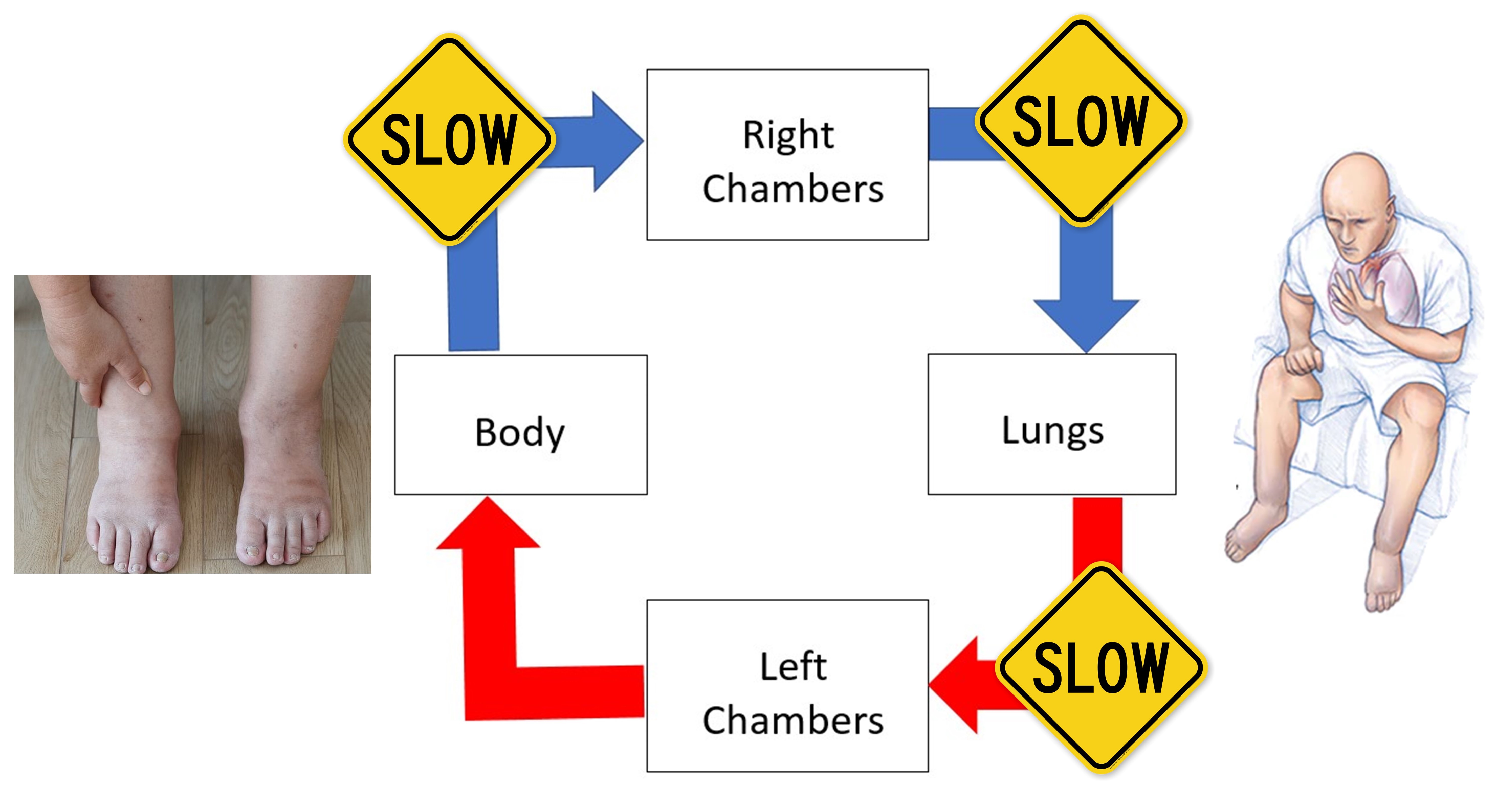
Cardiac amyloidosis occurs when an abnormal protein — called amyloid — builds up in the heart tissue. This buildup makes it difficult for your heart to relax completely in between heart beats, making the heart stiff. This stops blood from getting into the heart. There is a “traffic-jam” of blood waiting to enter the heart all across the body, causing congestion. The congestion causes “leakage of fluid” from the blood into the tissues- manifesting as fluid in lungs, legs, various internal organs and elsewhere. The common symptoms are shortness of breath, swelling in legs, lethargy and loss of appetite. This is a progressive disease and an increasingly recognized cause of heart failure.

Read further to understand this in detail.
Normal heart:
Heart is supposed to relax completely in-between heart beats. This allows the blood to enter the heart chambers, like a balloon blowing up with air (figure 1).
The relaxation of the heart is an equally important requirement to keep the blood moving forward in the continuous circuit around the body.
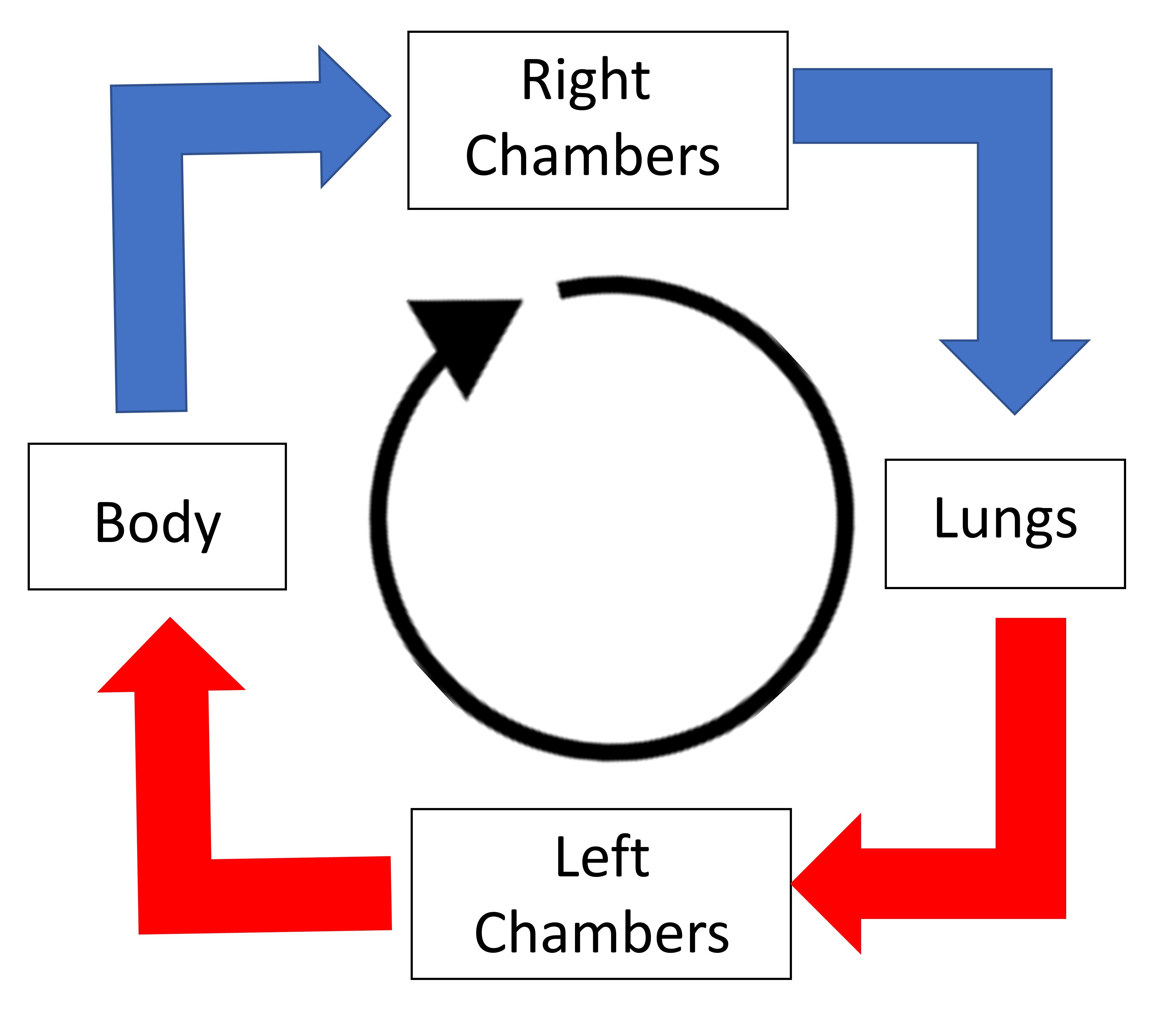
The circuit around the heart:
This has been explained in details elsewhere
In brief, the blood goes around the body as shown in figure 2. Here, the left and right chambers are the chambers of the heart.

The issue with cardiac amyloidosis:
The essential problem is deposition of abnormal and sticky proteins (amyloid) in various organs which interfere with the function of that organ. When the deposition occurs in the heart, this is called cardiac amyloidosis.
Two main types of cardiac amyloidosis:
There are two main types of cardiac amyloidosis:
- Transthyretin amyloidosis (ATTR amyloidosis): This type results from mutated deposits of transthyretin, a protein made by the liver. The two subtypes of ATTR are:
- Wild-type amyloidosis: Usually affects people in their 60s or older
- Hereditary amyloidosis: Runs in families and typically affects people in their 40s or older
- Light chain amyloidosis (AL amyloidosis): This type of amyloidosis is associated with blood cancers like multiple myeloma. It is not a type of cancer, but it is treated with chemotherapy.
The type of amyloid protein deposited in the above two types of cardiac amyloidosis is completely different and formed by different mechanisms, however the essential problem is the same- abnormal protein deposition.
AL amyloidosis
In AL amyloidosis, excessive abnormal antibodies are being formed by abnormal antibody-forming cells. The antibody forming factory has malfunctioned and starts manufacturing multiple batches of unusable antibodies. These antibodies belong to the same mould and are being manufactured in large quantities (figure 3).

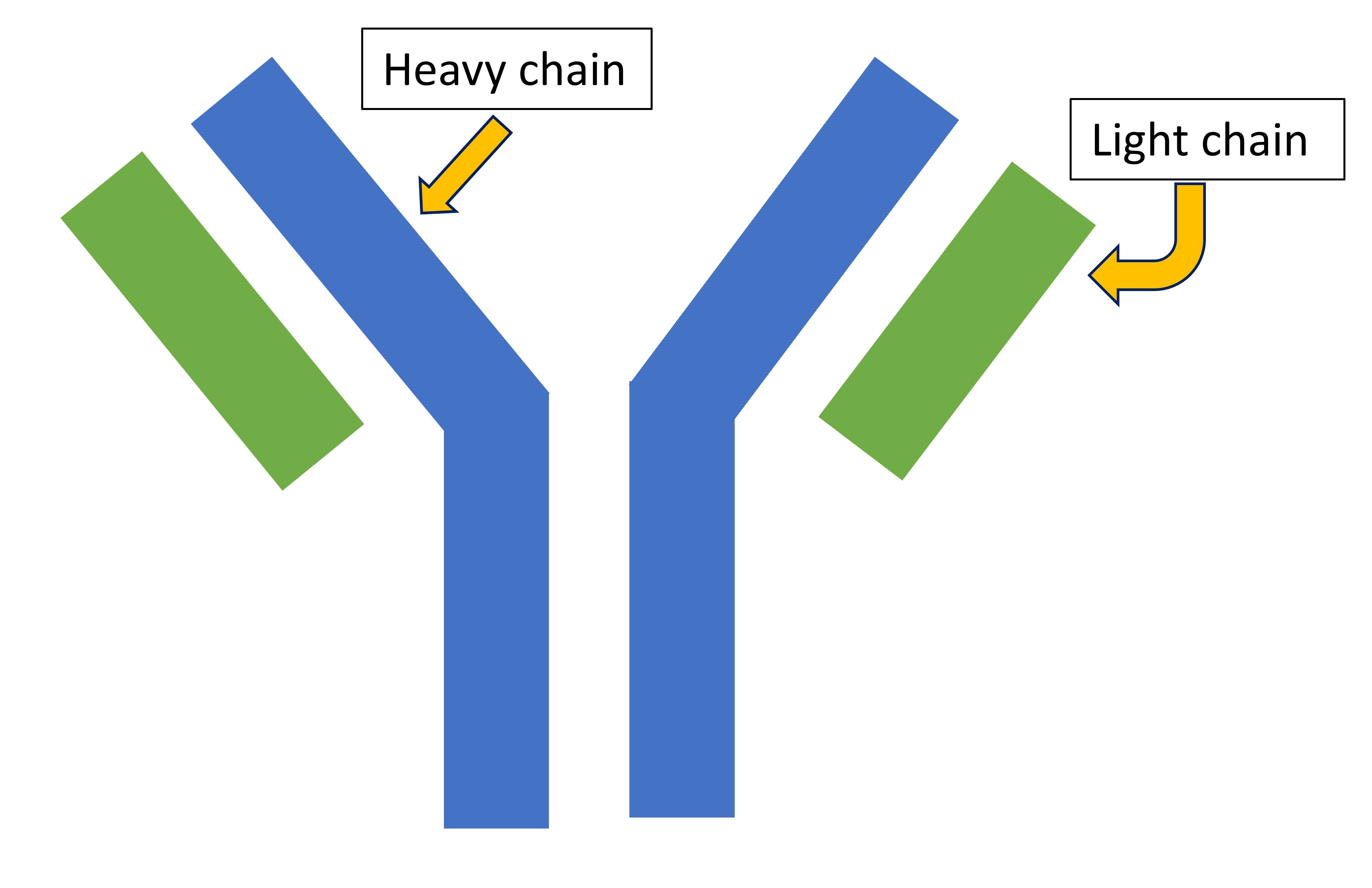
Antibodies are made of heavy chains and light chains (figure 4)

Light chains come in two forms – kappa and lambda. Each antibody is either made of kappa or lambda only
In AL amyloidosis, the abnormal antibody is made of only one type of light chain- either kappa or lambda. With the excessive antibody formation, the associated light chain (kappa or lambda) is formed in excessive amounts. The light chains may combine together, like clumps of hair, to form larger clumps called AL amyloid. These then accumulate in various organs and interfere with their function.
ATTR amyloidosis
Transthyretin (TTR) is a protein formed in the liver and functions to transport thyroid hormone and vitamin A around the body, like Uber.
In some patients, due to a genetic cause (hereditary form) or an unknown reason (wild-type), the transthyretin molecule formed by the liver is unstable and dismantles readily, like tires coming apart in a car. The dismantled molecule then gets misfolded.
The misfolded molecules cannot be broken down by the body and starts aggregating in the body, like plastic aggregating in the sea (figure 5).

Over time, these abnormally folded TTR molecules aggregate to form amyloid proteins, which accumulate in various organs.
Cardiac amyloidosis
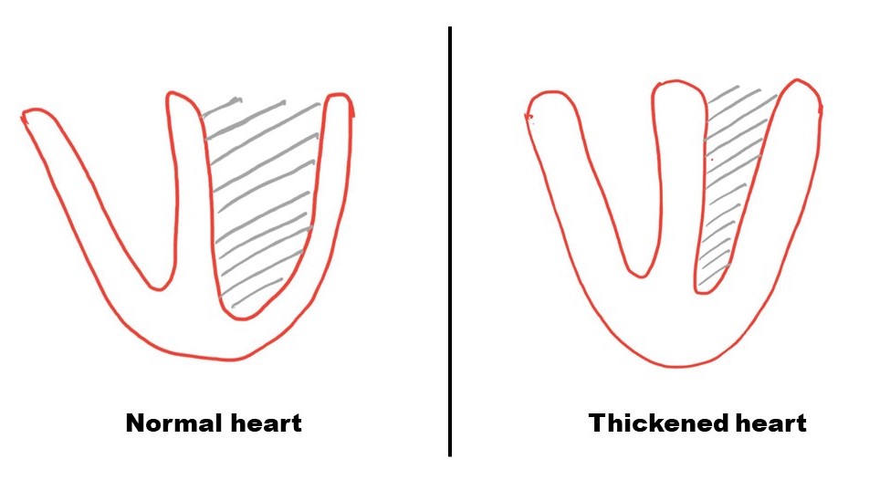
The amyloid proteins accumulate over time in between the heart muscles, making the heart stiff and thick. Like a balloon becoming stiff like a car tire.
The thickening of the heart muscles encroaches on the blood containing cavity, reducing the amount of blood it can contain (figure 6).


Not only does the heart get thickened but it also cannot expand completely in between the heart beats, making it difficult for the blood to get into the heart (figure 7, compare to figure 1).
.
.
As a consequence, there is a traffic-jam of blood all over the body, called “congestive heart failure”. The blood leaks fluid into the tissues and organs causing swelling and interference with the function of those organs (figure 8). This manifests as various symptoms including swelling in legs, shortness of breath, inability to sleep flat, lethargy, loss of appetite, kidney and liver failure.

For further information on the disease and the available treatment, refer to the UKY website and also listen to the UKHealthcare podcast on this topic.
All opinions expressed here are those of the author and not of the employer. Information provided here is for medical education only. It is not intended as and does not substitute for medical advice. The discussion is not all-inclusive or exhaustive and individual patient findings may vary. By use of this page, you agree to the disclaimer.