
Simple explanation of complex heart anatomy
Anatomy of the heart:
Blood pumping function (see figures):
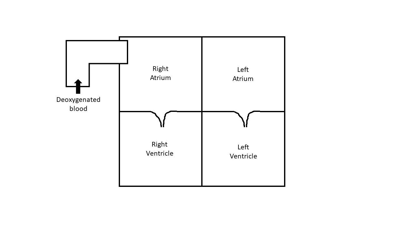
Heart has four chambers, like four adjoining rooms connected through doors. These doors or valves allow blood to flow in one direction only. The right sided rooms receive deoxygenated blood from the body through the veins. This blood first enters the right upper room where the blood is stored until the right lower room becomes available to receive blood (figure 1).
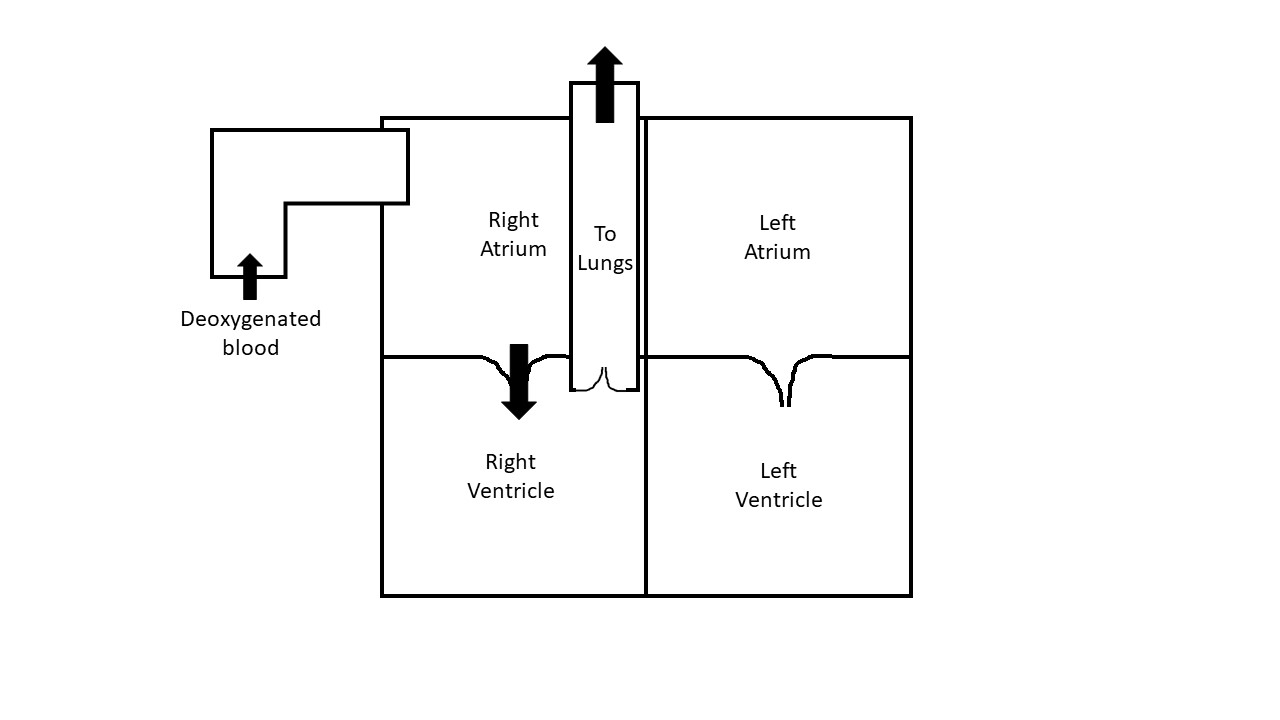
The blood then enters the right lower room through a door called the tricuspid valve. Thereafter, the blood is pumped into the lungs from the right lower room to get oxygenated (figure 2). This constitutes one heart-beat.
Simultaneously, the oxygenated blood from the lungs first enters the left upper room and then into the left lower room through a door called the mitral valve (figure 3).
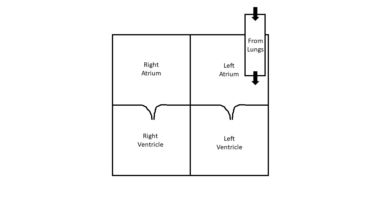
Figure 3: Simultaneously the oxygenated blood enters the left upper chamber
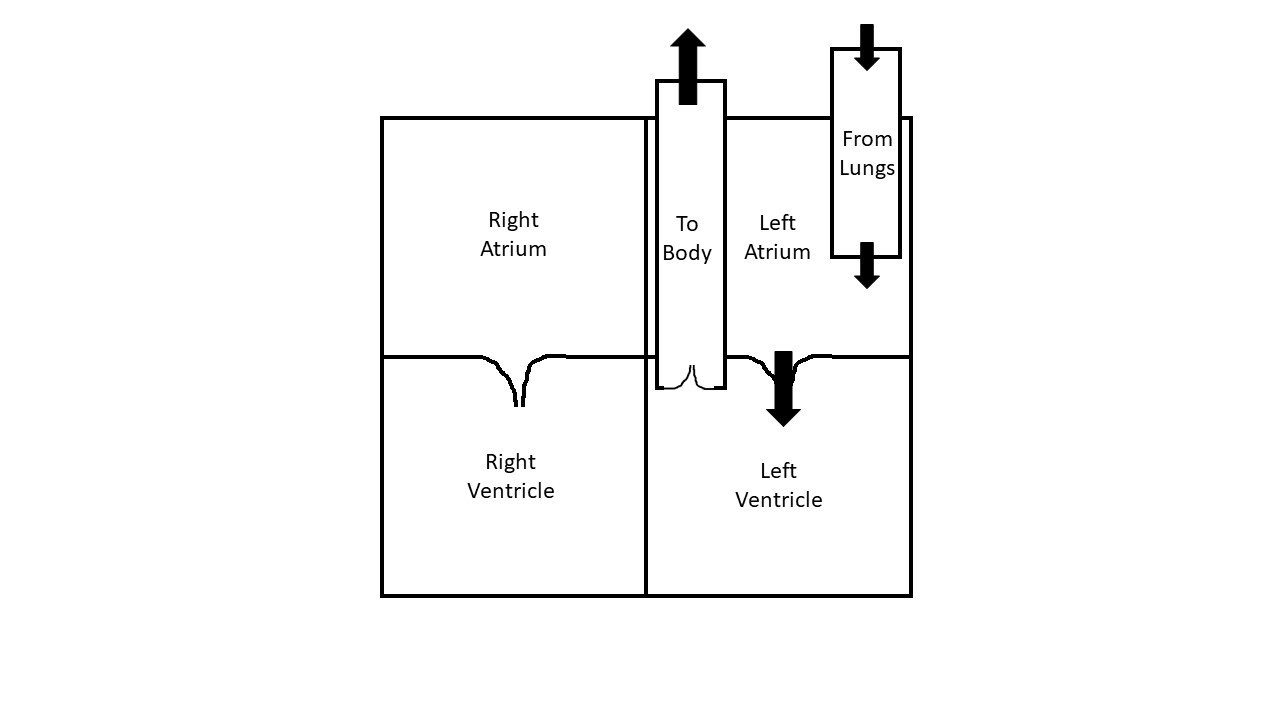
The blood is then pumped to the rest of the body through a door called the aortic valve (figure 4).
All these activities are happening simultaneously in the right and the left rooms independently. Normally there is no connection or door between the right and left rooms.
The big artery leaving the left lower room, carrying the oxygenated blood to the body, is called the aorta. Similarly, the big artery carrying deoxygenated blood to the lungs is called the pulmonary artery.
Normally, about 55-60% of the blood within the lower rooms is pumped forward during each squeeze.
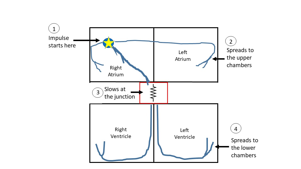
Anatomy of the electrical wiring of the heart (figure 5):
A heart to function normally needs to have a rhythm. The rooms need to beat in a rhythm, like symphony in an orchestra, to allow blood to flow between rooms smoothly. Just like the pattern of decoration lights in Christmas or Diwali, there is a progression of blood from one chamber to the next. For the automobile enthusiasts, the heart is like a four-stroke engine, where all the strokes need to happen sequentially for the crankshaft to rotate in one direction.
The sequence of these movements and the frequency (fast or slow) is decided by specialized cell aggregates contained within the right upper room, called the sinus node. Just like the drummer in a snake boat from Kerala or the dragon boat from China, who drums a rhythm and the paddlers paddle to this rhythm to maintain synchronicity. The sinus node is the drummer of the heart under normal circumstances. The electrical impulses then spread to the upper rooms first through specialized cells which function like electrical wires installed in your house. Consequently, the upper rooms (right and left) are the first to contract.
The electrical impulse then reaches a junction point, a border-crossing of sorts, in an effort to reach the lower rooms. This junction is called the atrioventricular or AV node. In normal conditions, AV node is the only way electrical impulse could travel from the upper to lower rooms. As in any border-crossing, the flow of traffic (electrical impulse in this case) is slowed down, so that only a certain number of cars (impulses) can go through, in a given time. This slowing also makes sure that the upper and lower rooms do not contract simultaneously.
Once the electrical impulses pass through the node successfully, they enter a highway headed either to the right or the left lower room. Thus, both the left and right lower rooms get activated quite rapidly and simultaneously, resulting in their contraction. The blood is pumped forward. This cycle repeats several times a minute, the frequency decided by the sinus node. Under stressful situations, such as exercise the sinus node responds to the increased demand in the body by increasing the heart rate. On the other hand, during restful situations such as sleeping, sinus node goes relatively slow.
Anatomy of blood supply to the heart:
Heart is literally filled with blood all the time, but the blood supply to the heart, through which it gets nutrition and oxygen, is provided through arteries. It is quite paradoxical that the heart is unable to get its requirements directly through the blood in its chambers but must rely on the blood supply through arteries like any other organ.
The arteries supplying the heart are called coronary arteries which arise from the aorta. Normally there are three main coronary arteries. Left anterior descending artery, notoriously called the ‘widow-maker’ is the largest and supplies the front of the heart. The other two arteries – left circumflex and the right coronary arteries, surround the heart in the middle and supply the left, right and posterior sides of the heart. All three arteries divide into various branches, getting smaller as they go further in their course. These three arteries are assigned the responsibility of supplying the entire real estate of the heart.
Anatomy of nerve supply to the heart:
The heart is like a tethered horse which must obey the orders from its master, the nervous system. The nervous system controls the heart through two opposing forces, like yin and yang. While one system speeds up the heart, makes the ‘border crossing’ faster and the heart to squeeze stronger; the other system does the opposite. These forces are required to meet the changing requirements of the body – to increase the blood supply when the requirement goes up and then slow down once the requirement ceases to exist. This explains why your heart speeds up during exercise or with certain emotions.
Miscellaneous:
Heart is contained within a bag called the pericardium. The pericardium has two layers which slip on each other and are lubricated by a thin layer of fluid, which in turn lubricates the motion of the heart within the body. The heart is located on the left side of the chest and is in close proximity to the left lung towards its left and the esophagus behind. Pain originating from these organs can therefore mimic each other.
All opinions expressed here are those of the author and not of the employer. Information provided here is for medical education only. It is not intended as and does not substitute for medical advice.
Follow on twitter.
