Implanted defibrillator or a ‘shock box’
A defibrillator is a device implanted to monitor the heart rhythm and give a shock when necessary. There are some reasons when this may be necessary.
What is happening in the heart?
Heart is a big net of electrical wiring, with a constant flow of organized electrical activity. This electrical activity gives the heart muscles the stimulus to contract in a sequential manner. Just like the pattern in Christmas lights or a Mexican wave in a stadium, the electrical stimulus moves a wave of contraction which pumps the blood out.
Occasionally some events may cause unusual electrical activity which could overwhelm the entire wiring system within the heart. If a zone of muscles is starved from oxygen (resulting from a blockage in blood supply like a heart attack), these muscle cells may act up electrically. Just like salary-starved laborers could start a strike and bring the company to a stop. This electrical storm, called ventricular tachycardia or fibrillation, moves so fast that it brings the heart to a standstill.
Unusual electrical activity may also happen around a scar in the heart such as after a heart attack. The boundary between the scar and normal muscles is electrically vulnerable for such sparks. Like an active border between enemy countries, cross-fires are common. There are also other reasons for these overwhelming electrical events including weakened heart pump, certain hereditary conditions, excessive electrolyte problems such as very high blood potassium levels, inflammation of heart tissue, certain medications and illicit drugs.
Irrespective of the cause, the best way to end this hurricane is to stop all activity on its tracks. Imagine a stadium full of people talking, shouting and playing music. The noise is deafening. A sound over the loudspeaker draws everyone’s attention and there is silence. An electrical shock works like the sound over the speaker. It attempts to stop the electrical chaos in the heart and allow organization to return. As you can imagine, shock only works in conditions resulting from an electrical chaos. It does not work to ‘charge’ the heart in an electrical standstill as they show in the movies.
The role of a defibrillator:
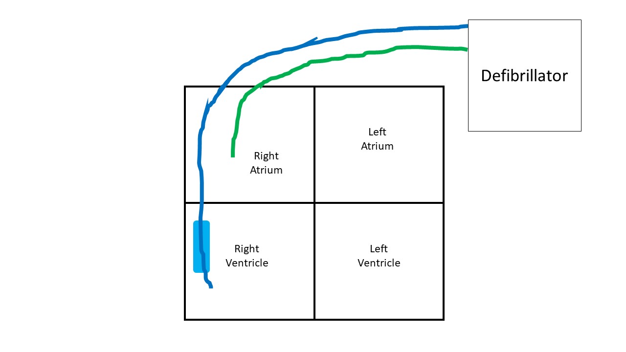
Just like a pacemaker, an implanted defibrillator is a generator implanted over the chest wall, underneath the skin. Wires or leads extend from the generator, enter a vein in the shoulder and travel to the heart. The most common defibrillator sends two leads to the heart (dual chamber). One of the lead goes into the right upper chamber and the second into the right lower chamber (figure 1).

The implanted defibrillator monitors the rhythm, like a personal CCTV camera keeping a close look on the heart. Based on the programming of the device, if the electrical activity gets faster than the programmed number, the device attempts to shock and bring back organized activity. The shock may be repeated if the initial attempt is unsuccessful.
The defibrillator also doubles up as a pacemaker and can do all its functions as well.
Defibrillator care after placement:
It is recommended to not lift the arm above the head for 2 weeks. A splint may be provided to keep the arm immobilized. This is to prevent the suture site from opening. Some swelling around the defibrillator site immediately after placement is expected, however it is unusual for the swelling to continue growing, appear excessively red, warm, firm or be painful. Drainage or bleeding from the site is also usually abnormal. The defibrillator should be completely underneath the skin and at no point should any part stick out.
Shortness of breath, chest pain and dizziness are concerning symptoms after implantation and need immediate medical attention.
MRI compatibility:
Many of the old defibrillators are not FDA-approved to be MRI compatible. Some of the new defibrillators have been tested and approved as MRI-compatible, check with your physician. Also the radiologists need to be made aware of the defibrillator, and some programming changes may be necessary prior to an MRI.
On occasions, the defibrillator leads may be abandoned within the heart, when removing them is riskier than any benefit. This is especially true for leads placed more than a few months ago, due to scar formation around the leads. In such cases, MRI may cause overheating of these abandoned leads. The presence of abandoned leads should be notified to the physician prior to an MRI.
At the airport:
Inform the TSA officials at the airport of the presence of a defibrillator. A pat-down may be requested.
Prior to a scheduled surgery:
The presence of a defibrillator needs to be notified to the surgeon. The surgeon may consult with the company representative, who may ask for a visit before and/or after the surgery to check the defibrillator.
Troubleshooting:
If the implanted defibrillator makes noise, this needs to get checked. It usually signifies that the battery is up for renewal, but there are other conditions when it may make a noise. During the battery replacement procedure, the generator on the chest wall is swapped with a new one. It is essential to get the defibrillator checked at least once a year. If the defibrillator delivers a shock, then a visit to the closest emergency department is necessary.
Drainage or infection may require the defibrillator to be removed completely.
Some facts:
The defibrillator is a machine and works based on certain algorithms set while programming. Consequently, it can make mistakes at times and a shock may be delivered inappropriately. This is fortunately quite rare and usually remediable.
The shock delivering function of a defibrillator can be turned off if the patient wishes so.
A defibrillator can really save lives.
All opinions expressed here are those of the author and not of the employer. Information provided here is for medical education only. It is not intended as and does not substitute for medical advice.
Follow on twitter.
