What is an ECG / EKG?
Have you ever seen a doctor read the squiggly lines on the ECG and give you a diagnosis? Have you ever wondered what is in those squiggly lines that made the doctor exclaim- ‘This is normal’ or ‘You are having a heart attack!’. The description below is for a standard 12-lead ECG recorded at regular speed with standard lead placement.
ECG is like sheet music; each notation stands for a key that represents the time when the note should be played. ECG is also a moving paper which recorded the electrical activity in the heart over time (usually 10 seconds).
While getting an ECG, a physician/nurse/technician places ‘leads’ on the chest and limbs. These record the electrical activity within the heart. This is of course at a very low amplitude (usually ~1-2 millivolt). The leads are placed methodically at various pre-decided points on the body and each record the electrical changes differently (figure 1).
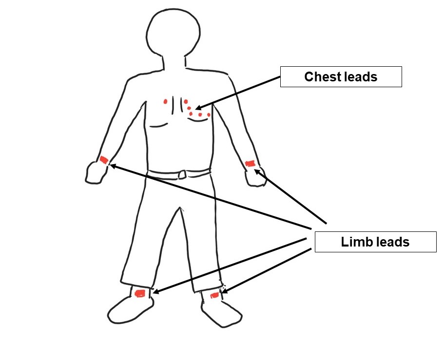
Think of leads like many windows in a room full of paintings. A person looking into the room from outside may see each of the paintings differently depending on which window she/he looks from. An art connoisseur can appreciate the Mona Lisa from various angles; up, down and the sides. The ECG leads are the ‘heart’ connoisseur.
In simple terms, electrical current swishes back and forth within the heart with each heart-beat. This electrical activity is recorded by a particular lead as an upstroke when the electrical current travels ‘towards’ the lead, or vice versa. Therein lies the description of ECG. Each lead picks up the same electrical activity differently based on how it is looking at it (like the window analogy above).
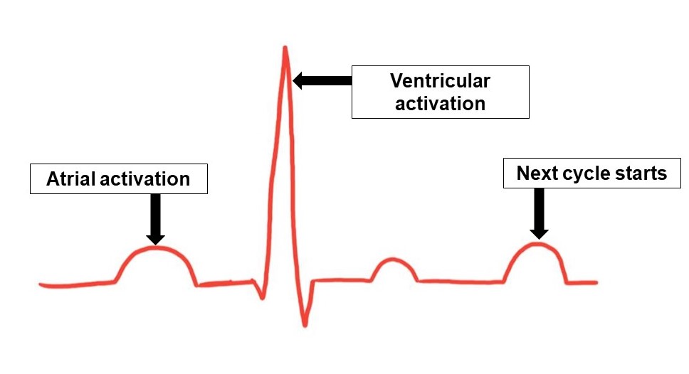
As explained in the basic heart description page, the electrical activity usually first starts in the right upper chambers and then spreads to both the upper chambers (figure 2).
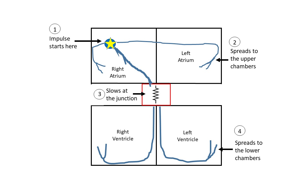
Consequently, the first wave on the ECG is that of the atrial (upper chamber) activation. The electrical activity then travels towards the lower chambers and encounters a ‘border-crossing’ of sorts where the electrical current gets slowed (called the AV node), depicted as number 3 on the figure above. Once the current passes through this border-crossing, the current spreads through both the lower chambers simultaneously, in normal circumstances. So, the next wave on the ECG is that of the ventricular (lower chamber) activation (figure 3).

There are many factors that get recorded such as the heart rate, rhythm, strength of electrical activity, unexpected electrical activation such as atrial fibrillation or any evidence of injury or malfunction. Each of these possibilities create a recognizable pattern on the ECG which is beyond the scope of this discussion, but the reader is referred to the great resources online.
The ECG is an old discovery which continues to have application in modern medicine due to its simplicity, portability, speed and reliability. Certain ECG patterns are quite specific for certain life-threatening heart conditions while others need confirmation through other modes of diagnosis. Just like everything in medicine, ECG is not an exact science.
Follow on twitter.
One Reply to “What is an ECG / EKG?”
Comments are closed.